This 30-year-old lady, a nurse by profession working in a big Corporate Hospital of Siliguri from North Bengal, contacted us as she was incidentally diagnosed with a rare condition of pulmonary multifocal AVF for possible interventional treatment. She is married having 3-year-old kid as well but the condition was never diagnosed earlier. During one routine ward round while cheking Patient’s oxygen saturation, she tried it on herself as well and found to have very low oxygen saturation almost not compatible with existence of less than 80%. This was repeatedly measured and then HRCT thorax was performed which showed 2 possible large pulmonary AVF supplied by multiple feeders in the right lower lobe medial basal segment with large draining veins to right lower lobe pulmonary vein.
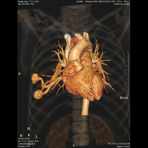
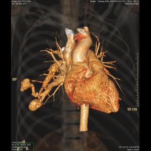
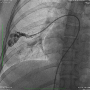
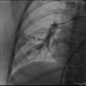
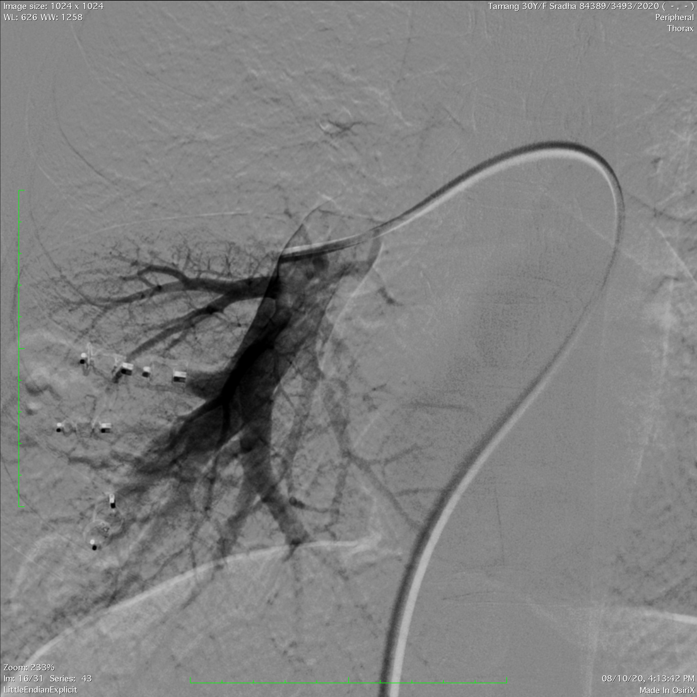
On presentation, a CTPA performed here which confirmed the diagnosis having two large AVF in the right lower lobe medial basal segment with upper one supplied by at least 3 feeders and lower one by two feeders, leading to large venous sacs and then to draining vein to right lower lobe. A transthoracic echocardiography with contrast showed moderate degree of shunt. She is suffering from chronic hypoxemia , she needed treatment to prevent any further complication especially like of brain abscess.
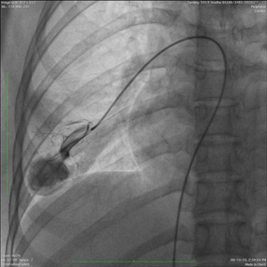
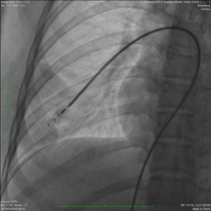
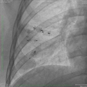
Via right trans femoral venous access, a 6-Fr.sheath was placed and using an Envoy guiding catheter over wire, all the feeders were selectively and Superselectively one by one cannulated and blocked using vascular plugs. Lower AVF was larger and supplied by a large main feeder which was peripherally divided into two before directly opening up in the dilated large venous sac, was occluded by a large plug. The upper one having 2 distinct feeders from same branch which was occluded selectively and peripherally using 2 plugs. On check injection, another smaller feeder was seen from the lower branch to the upper AVF which was also surperselectively cannulated and blocked peripherally. Check injection show complete occlusion of AVF with immediate contrast stasis in the venous sacs and the draining veins. This, however, can result in acute stroke if dislodges further, so patient was observed in ITU and was started on IV HEPARIN infusion for next 24 hours and discharged following day on dual antiplatelets for 1 month. Immediate post-procedure her oxygen saturation improved and recorded as 100% in both supine as well as standing positions.
This type for pulmonary AVF are usually part of a syndrome called Osler-Weber-Rendu syndrome but no other stigmata of any other disease process could be demonstrated.
This type of pulmonary AVF are quite difficult to treat and has to be dealt in very proper way to avoid any possibility of thromboembolic phenomena leading to cerebral stroke, as emboli from the pulmonary circulation can directly travel to the large intracranial vessels and occlude them. Non-treating them can result in similarly infection travelling from the lung directly into the brain parenchyma, lodging there and leading to brain abscess formation ( many patients present with them only). This is superimposed on chronic hypoxemia can spill significant danger for the patients in any compromised situation. Successful treatment requires proper planning and blocking the feeders in a way which minimally compromises the rest of the pulmonary circulation.